Performing the Sterilization Procedure
Explaining the Procedure
A woman who has chosen female sterilization needs to know what will happen during the procedure. The following description can help explain the procedure to her. Learning to perform female sterilization takes training and practice under direct supervision. Therefore, this description is a summary and not detailed instructions.
(The description below is for procedures done more than 6 weeks after childbirth. The procedure used up to 7 days after childbirth is slightly different.)
The Minilaparotomy Procedure
- The provider uses proper infection-prevention procedures at all times (see Infection Prevention in the Clinic).
- The provider performs a physical examination and a pelvic examination. The pelvic examination is to assess the condition and mobility of the uterus.
- The woman usually receives light sedation (with pills or into a vein) to relax her. She stays awake. Local anesthetic is injected above the pubic hair line.
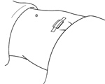
- The provider makes a small vertical incision (2–5 centimeters) in the anesthetized area. This usually causes little pain. (For women who have just given birth, the incision is made horizontally at the lower edge of the navel.)
- The provider inserts a special instrument (uterine elevator) into the vagina, through the cervix, and into the uterus to raise each of the 2 fallopian tubes so they are closer to the incision. This may cause discomfort.
 Each tube is tied and cut or else closed with a clip or ring.
 - The provider closes the incision with stitches and covers it with an adhesive bandage.
- The woman receives instructions on what to do after she leaves the clinic or hospital (see Explaining Self-Care for Female Sterilization). She usually can leave in a few hours.
The Laparoscopy Procedure
- The provider uses proper infection-prevention procedures at all times (see Infection Prevention in the Clinic).
- The provider performs a physical examination and a pelvic examination. The pelvic examination is to assess condition and mobility of the uterus.
- The woman usually receives light sedation (with pills or into a vein) to relax her. She stays awake. Local anesthetic is injected under her navel.
- The provider places a special needle into the woman's abdomen and, through the needle, inflates (insufflates) the abdomen with gas or air. This raises the wall of the abdomen away from the pelvic organs.
- The provider makes a small incision (about one centimeter) in the anesthetized area and inserts a laparoscope. A laparoscope is a long, thin tube containing lenses. Through the lenses the provider can see inside the body and find the 2 fallopian tubes.
- The provider inserts an instrument through the laparoscope (or, sometimes, through a second incision) to close off the fallopian tubes.
- Each tube is closed with a clip or a ring, or by electric current applied to block the tube (electrocoagulation).
- The provider then removes the instrument and laparoscope. The gas or air is let out of the woman’s abdomen. The provider closes the incision with stitches and covers it with an adhesive bandage.
- The woman receives instructions on what to do after she leaves the clinic or hospital (see Explaining Self-Care for Female Sterilization). She usually can leave in a few hours.
 Previous Chapter
  Previous Page Next Page
  Next Chapter